


Dental Radiography
Radiographs are valuable aids in the oral health care of infants, children, adolescents, and persons with special health care needs. They are used to diagnose oral diseases and to monitor dentofacial development and the progress of therapy. While children are more sensitive to x rays than adults because they are still growing, the amount of radiation from needed dental radiographs is extremely small, equivalent to a few hours of natural background radiation, which we have around us all the time. It is less radiation than they would receive if they made a trip to the mountains (higher background radiation at high altitudes) or flew in an airplane (increased cosmic radiation at flight levels). The minimal risk of the radiographs needs to be compared with the information obtainable that may be very important for your child's health.
Clinical situations for which radiographs may be indicated include but are not limited to:
- Deep carious lesions
- Mobility of teeth
- History of pain
- Growth abnormalities
- Traumatic injuries
- Unusual tooth morphology, calcification or color
- Malposed or clinically impacted teeth
- Unusual eruption, spacing or migration of teeth
- Developmental anomalies
- Unexplained absence of teeth
- Unexplained discoloration of teeth
- Orthodontic evaluation
- Presence of implants or evaluation for implant placement
- Evidence of swelling/fistula
- Problems of eruption
- Familial history of dental anomalies
- Large or deep restorations
- Clinical evidence of periodontal disease
- Unexplained sensitivity of teeth
Radiographs should only be used when the patient history and/or objective findings and symptoms lead to the conclusion that further useful information might be obtained. If a radiograph is not expected to change the diagnosis or treatment, or add other useful information, it should not be taken.
Commonly used radiographic techniques:
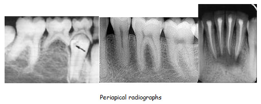
Periapical radiography: It is a one form of an intraoral techniques designed to show two to four teeth, the tissues around the apices and provides detailed information about the surrounding alveolar bone. The main clinical indications for periapical radiography include:
- Detection of apical infection/inflammation
- Assessment of the periodontal status
- After trauma to the teeth and associated alveolar bone
- Assessment of the presence and position ofunerupted teeth
- Assessment of root morphology before extractions
- During endodontics
- Preoperative assessment and postoperative appraisal of apical surgery
- Detailed evaluation of apical cysts and otherlesions within the alveolar bone
- Evaluation of implants postoperatively.
Panoramic radiography: Panoramic radiographs are to visualize the maxilla and the mandible including the teeth, maxillary sinuses, and nasal cavity. Panoramic films are extraoral films, in which the film is exposed while outside the patients' mouth.The principal advantages of the panoramic radiograph are the large area of coverage, the bilateral view of anatomy, low patient radiation dose, and it can be used on handicapped patients, children, and on those who are unable to open their mouths.

Bite-wing radiography: The bitewing view is taken to visualize the crowns of the posterior teeth and the height of the alveolar bone in relation to the cementoenamel junctions, which are the demarcation lines on the teeth which separate tooth crown from tooth root. Routine bitewing radiographs are commonly used to examine for interdental caries and recurrent caries under existing restorations.
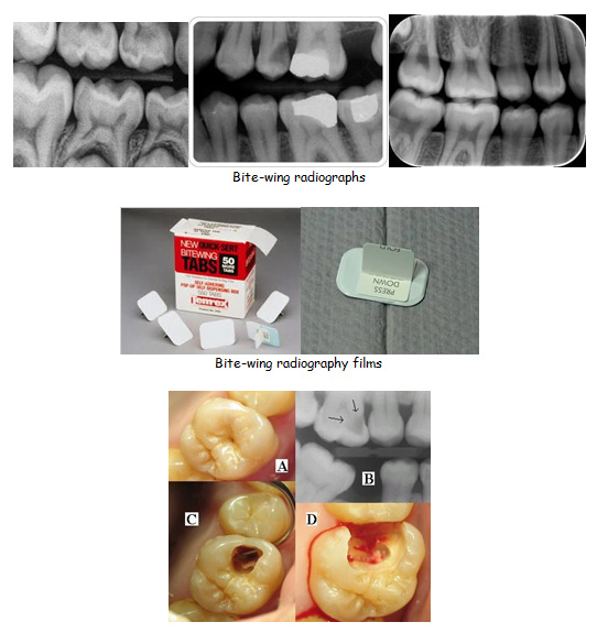
This preoperative photo of tooth #3, (A), reveals no clinically apparent decay other than a small spot within the central fossa. In fact, decay could not be detected with an explorer. Radiographic evaluation, (B), however, revealed an extensive region of demineralization within the dentin (arrows) of the mesial half of the tooth. When a bur was used to remove the occlusal enamel overlying the decay, (C), a large hollow was found within the crown and it was discovered that a hole in the side of the tooth large enough to allow the tip of the explorer to pass was contiguous with this hollow. After all of the decay had been removed, (D), the pulp chamber had been exposed and most of the mesial half of the crown was either missing or poorly supported.
Occlusal radiography: Used primarily for evaluation of supernumerary teeth, impacted canines, trauma to the anterior teeth. An occlusal radiograph will show:
- Presence or absence of primary teeth
- Presence or absence, and stage of development of permanent anterior teeth
- Presence of supernumery teeth
- Status of traumatized teeth
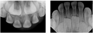
Anterior occlusal radiographs
Cephalometric radiography:It is a form of skull radiography used extensively in orthodontics to assess the relationships of teeth to the jaws to the rest of the facial skeleton. This diagnostic radiograph used primarily for orthodontic treatment planning, and is taken during the orthodontic records appointment. It is also used before and after the orthognathic surgery.
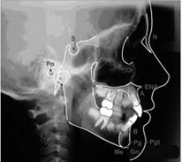
Cephalometric radiography
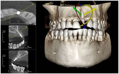
Computed tomography (CT): CT can be used to image the extent of pathologic conditions as well as help to unravel complex facial fractures. CT can also be used to assess the temporomandibular joints and the paranasal sinuses and for presurgical implant treatment planning.
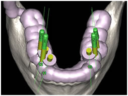
Cone beam computed tomography (CBCT): Cone beam computed tomography (CBCT) has added 3-dimensional capabilities that have many applications in dentistry. Specially designed CBCT scanners produce adequate imaging with a tenfold reduction in radiation than that of CT.
Digital radiography: It is a form of X-ray imaging, where digital X-ray sensors are used instead of traditional photographic film. Advantages include time efficiency through bypassing chemical processing and the ability to digitally transfer and enhance images. Also less radiation can be used to produce an image of similar contrast to conventional radiography. Instead of X-ray film, a small sensor unit sends pictures to a computer to be recorded and saved. Immediate image preview and availability; elimination of costly film processing steps; a wider dynamic range, which makes it to correct of over- and under-exposure images; as well as the ability to apply special image processing techniques that enhance overall display of the image are the advantages of digital radiography.
Sources
Serdar UYSAL Turkiye Klinikleri J Dental Sci-Special Topics 2010;1(2):36-43
http://jada.ada.org/content/135/10/1437.full
http://www1.umn.edu/dental/courses/dent_5501/till_handout02.pdf
Miles DA, Parks ET. Radiographic techniques. In: Dean JA, Avery DR, McDonald RE. Dentistry for the child and adolescent. St. Louis, Mosby Elsevier, 9th ed., 2011, p:47-63.
http://www.aapd.org/media/policies_guidelines/e_radiographs.pdf 2011-2012
http://v5.books.elsevier.com/bookscat/samples/9780443102134/9780443102134.pdf
Parks ET. Computed tomography applications for dentistry.Dent Clin North Am. 2000 Apr;44(2):371-94.
http://www.milesofsmilesdental.net/783/using-cone-beam-ct-cbct-technology-to-plan-complex-implant-placement-in-the-maxilla/
http://www.scribd.com/doc/21866130/Cephalometric-Radiography
http://en.wikipedia.org/wiki/Dental_radiography
http://en.wikipedia.org/wiki/Digital_radiography